How To Detect Microvesicles, Microparticles, And Ectosomes By Flow Cytometry

There seems to be a big fuss about small flow these days.

What is small flow?

Microvescles, microparticles, ectosomes—the terminology is all over the place but one thing is certain…

There is a need for flow cytometric analysis of extracellular vesicles.

To avoid ambiguity in this post, the population of extracellular vesicles discussed here shall be called microvesicles.

These small, membrane bound fractions of cells released during activation and apoptosis are in the 0.1 to 1 micron diameter size range (or there about depending on which article you read).

Released in vast quantities and carrying a variety of traits from the originating cell these microvesicles are known to be involved in a wide range of processed from stem cell renewal and tumor metastases through to coagulation and inflammation, just to name a few!

They key is that microvesicles have a huge potential to be important biomarkers in early disease detection and/or treatment progression.

What’s Down There? Debris Or Microvesicles?

Microvesicles have been known about for a long time, being dismissed by the regular flow cytometrist as debris or platelet dust and quickly gated out of any subsequent analysis.

microvesicle experiment by flow cytometry | Expert Cytometry | microparticle analysis

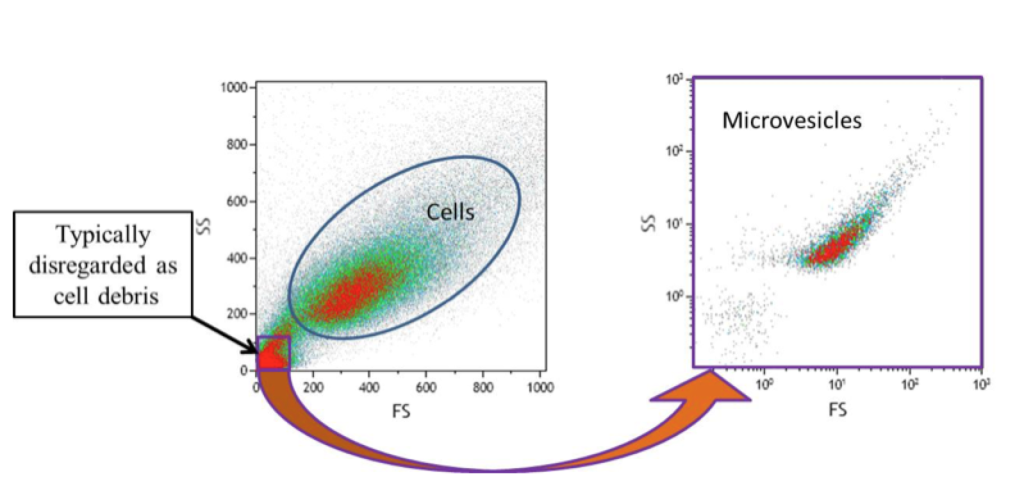
There is increasing interest in seeing what is down there. As in, what is down in the lower left-hand corner of the above figure.

Is it debris? Is it platelet dust? Is it a population of microvesicles?

Or is it all three?

The above figure is a representation of what we are missing when we ignore events in that range.

It’s important to note that the sample on the right in the above figure was further processed before re-analysis in the microvesicle range.

4 Microvesicle Flow Cytometry Mistakes To Avoid

Microvesicles originate from cells and have the same analysis requirements as cells.

For these and other reasons, flow cytometry is a popular choice for microvesicle analysis. However, there are pitfalls with small particle flow cytometry that have led to many conflicting publications.

The only way to avoid these mistakes is to first identify them and then take measures to prevent them.

The following are 4 common mistakes researchers make when preparing microvesicle flow cytometry experiments, as well as how to prevent these mistakes…

1. Not accounting for flow cytometry protocol discrepancies.

Before getting started with your microvesicle experiment, you must take into account pre-analytical variations. When the particles are this small, even the slightest protocol variations can affect your experiment.

Centrifugation speed and duration, collection technique, storage, staining protocols (in particular antibody panels and titration levels) are just a few of the things you should consider.

Minor differences in these protocol steps can lead to huge discrepancies in downstream analysis.

2. Using beads to define microvesicle size ranges.

When setting up for the analysis of microvesicles, many people choose flow cytometry beads to define a size range.

The problem here is, in terms of standards, the different refractive index between beads and microvesicles becomes troublesome.

For example, a polystyrene bead may have a refractive index of about 1.59 whereas while the refractive index of many microvesilces are around 1.39 (for reference, water is 1.33).

Beads scatter a lot more light compared to microvesicles of the same diameter, reportedly up to 100 times!

Here’s the lesson—just because you can see a 0.2 micron diameter bead by flow does not mean you can see a 0.2 micron diameter microvesicle by flow.

3. Failing to acknowledge the possibility of unseen information.

Microvesicles are between 0.1 – 1 micron diameters in size. Though most fall in the smaller end of that range.

Commercial cytometers can struggle to detect light from a 0.2 micron diameter bead. As discussed in point #2 above, beads scatter a LOT more light than microvesicles.

With this in mind you have to ask yourself, how many microvesicles can I actually see by flow cytometry?

According to many flow cytometrists—not a lot.

When analyzing microvesicles by conventional flow cytometry, it’s worth considering the massive amount of information that goes unseen.

Don’t assume you’re seeing all the microvesicles in your experiment. Instead, constantly ask yourself, “What am I NOT seeing?”

As techniques improve, you’ll be able to see more and more information and continue to get a clearer picture of what’s really going on in your microvesicle flow cytometry experiments.

4. Failing to account for coincident events.

Accounting for coincidence events is important to every flow cytometry experiment, including experiments measuring and reporting microvesicles.

This is especially true if you’re hoping to publish the flow cytometry data you obtain.

The problem here is that conventional doublet discrimination in this microvesicle size ranges is exceedingly difficult.

A single event, or dot, on a flow cytometry plot may actually be many, many microvesicles passing through the laser beam at the same time. The only way to know for sure is to check.

One way to check your microvesicle analysis is with serial dilutions.

If you’re able to see a stable fluorescent signal with dilutions resulting in a lower event count, you can gain more certainty over the assumption that you’re analyzing (mostly) single events.

The analysis of microvesicles by flow cytometry is not easy. However, until better instrumentation is developed, in wide use, and shown to be more accurate, using conventional flow cytometry to detect and measure microvesicles is of great value. While a multi-platform strategy continues to be the wisest approach, much information on microvesicles can be gained by conventional flow cytometry.

To learn more about detecting microvesicles by flow cytometry and to get access to all of our advanced materials including 20 training videos, presentations, workbooks, and private group membership, get on the Flow Cytometry Mastery Class wait list.

Join Expert Cytometry's Mastery Class

ABOUT TIM BUSHNELL, PHD

Tim Bushnell holds a PhD in Biology from the Rensselaer Polytechnic Institute. He is a co-founder of—and didactic mind behind—ExCyte, the world’s leading flow cytometry training company, which organization boasts a veritable library of in-the-lab resources on sequencing, microscopy, and related topics in the life sciences.

Tim Bushnell, PhD

Similar Articles

The Power Of Spectral Viewers And Their Use In Full Spectrum Flow Cytometry

The Power Of Spectral Viewers And Their Use In Full Spectrum Flow Cytometry

By: Tim Bushnell, PhD

What photon from yonder fluorochrome breaks?  It is … umm… hmmm. Let me see. Excitation off a 561 nm laser, with an emission maximum of 692 nm. I’m sure if Shakespeare was a flow cytometrist, he might have written that very scene. But the play is lost in time. However, since the protagonist had difficulty determining what fluorochrome was emitting photons, let’s consider how this could be figured out. In my opinion, one of the handiest flow cytometry tools is the spectral viewer. This tool helps visualize the excitation and emission profile of different fluorochromes, as well as allowing you…

Common Numbers-Based Questions I Get As A Flow Cytometry Core Manager And How To Answer Them

Common Numbers-Based Questions I Get As A Flow Cytometry Core Manager And How To Answer Them

By: Tim Bushnell, PhD

Numbers are all around us.  My personal favorite is ≅1.618 aka ɸ aka ‘the golden ratio’.  It’s found throughout history, where it has influenced architects and artists. We see it in nature, in plants, and it is used in movies to frame shots. It can be approximated by the Fibonacci sequence (another math favorite of mine). However, I have not worked out how to apply this to flow cytometry.  That doesn’t mean numbers aren’t important in flow cytometry. They are central to everything we do, and in this blog, I’m going to flit around numbers-based questions that I have received…

3 Must-Have High-Dimensional Flow Cytometry Controls

3 Must-Have High-Dimensional Flow Cytometry Controls

By: Tim Bushnell, PhD

Developments such as the recent upgrade to the Cytobank analysis platform and the creation of new packages such as Immunocluster are reducing the computational expertise needed to work with high-dimensional flow cytometry datasets. Whether you are a researcher in academia, industry, or government, you may want to take advantage of the reduced barrier to entry to apply high-dimensional flow cytometry in your work. However, you’ll need the right experimental design to access the new transformative insights available through these approaches and avoid wasting the considerable time and money required for performing them. As with all experiments, a good design begins…

The Fluorochrome Less Excited: How To Build A Flow Cytometry Antibody Panel

The Fluorochrome Less Excited: How To Build A Flow Cytometry Antibody Panel

By: Tim Bushnell, PhD

Fluorochrome, antibodies and detectors are important. The journey of a thousand cells starts with a good fluorescent panel. The polychromatic panel is the combination of antibodies and fluorochromes. These will be used during the experiment to answer the biological question of interest. When you only need a few targets, the creation of the panel is relatively straightforward. It’s only when you start to get into more complex panels with multiple fluorochromes that overlap in excitation and emission gets more interesting.  FLUOROCHROMES Both full spectrum and traditional fluorescent flow cytometry rely on measuring the emission of the fluorochromes that are attached…

Flow Cytometry Year in Review: Key Changes To Know

Flow Cytometry Year in Review: Key Changes To Know

By: Meerambika Mishra

Here we are, at the end of an eventful year 2021. But with the promise of a new year 2022 to come. It has been a long year, filled with ups and downs. It is always good to reflect on the past year as we move to the future.  In Memoriam Sir Isaac Newton wrote “If I have seen further, it is by standing upon the shoulders of giants.” In the past year, we have lost some giants of our field including Zbigniew Darzynkiwicz, who contributed much in the areas of cell cycle analysis and apoptosis. Howard Shapiro, known for…

What Star Trek Taught Me About Flow Cytometry

What Star Trek Taught Me About Flow Cytometry

By: Tim Bushnell, PhD

It is no secret that I am a very big fan of the Star Trek franchise. There are many good episodes and lessons explored in the 813+ episodes, 12 movies (and counting). Don’t worry, this blog is not going to review all 813, or even 5 of them. Instead, some of the lessons I have taken away from the show that have applicability to science and flow cytometry.  “Darmok and Jalad at Tanagra.”  (ST:TNG season 5, episode 2) This is probably one of my favorite episodes, which involves Picard and an alien trying to establish a common ground and learn…

5 Flow Cytometry Strategies That Sun Tzu Taught Me

5 Flow Cytometry Strategies That Sun Tzu Taught Me

By: Tim Bushnell, PhD

Sun Tzu was a Chinese general and philosopher. His most famous writing is ‘The Art of War’, and has been studied by generals and CEOs, to glean ideas and strategies to help their missions. I was recently rereading this work and thought to myself if any of Sun Tzu’s lessons could apply to flow cytometry.  So I have identified 5 points that I think lend themselves to thinking about flow cytometry.  “Quickness is the essence of the war.” In flow cytometry, speed is of the essence. The longer the cells are out of their natural environment, the less happy they…

A Basic Guide To Flow Cytometry (3 Foundational Concepts)

A Basic Guide To Flow Cytometry (3 Foundational Concepts)

By: Meerambika Mishra

Mastering foundational concepts are imperative for successfully using any technique or system.  Robert Heinlein introduced the term ‘Grok’  in his novel Stranger in a Strange Land. Ever since then it has made its way into popular culture. To Grok something is to understand it intuitively, fully. As a cytometrist, there are several key concepts that you must grok to be successful in your career. These foundational concepts are the key tools that we use day in and day out to identify and characterize our cells of interest.  Cells Flow cytometry measures biological processes at the whole cell level. To do…

How To Do Variant Calling From RNASeq NGS Data

How To Do Variant Calling From RNASeq NGS Data

By: Deepak Kumar, PhD

Developing variant calling and analysis pipelines for NGS sequenced data have become a norm in clinical labs. These pipelines include a strategic integration of several tools and techniques to identify molecular and structural variants. That eventually helps in the apt variant annotation and interpretation. This blog will delve into the concepts and intricacies of developing a “variant calling” pipeline using GATK. “Variant calling” can also be performed using tools other than GATK, such as FREEBAYES and SAMTOOLS.  In this blog, I will walk you through variant calling methods on Illumina germline RNASeq data. In the steps, wherever required, I will…

Top Industry Career eBooks

Get the Advanced Microscopy eBook

Get the Advanced Microscopy eBook

Heather Brown-Harding, PhD

Learn the best practices and advanced techniques across the diverse fields of microscopy, including instrumentation, experimental setup, image analysis, figure preparation, and more.

Get The Free Modern Flow Cytometry eBook

Get The Free Modern Flow Cytometry eBook

Tim Bushnell, PhD

Learn the best practices of flow cytometry experimentation, data analysis, figure preparation, antibody panel design, instrumentation and more.

Get The Free 4-10 Compensation eBook

Get The Free 4-10 Compensation eBook

Tim Bushnell, PhD

Advanced 4-10 Color Compensation, Learn strategies for designing advanced antibody compensation panels and how to use your compensation matrix to analyze your experimental data.