Optimizing Flow Cytometry Experiments – Part 2 How To Block Samples (Sample Blocking)

In my previous blog on experimental optimization, we discussed the idea of identifying the best antibody concentration for staining the cells. We did this through a process called titration, which focuses on finding the best signal-to-noise ratio at the lowest antibody concentration. In this blog we will deal with sample blocking
As a reminder, there are two other major binding concerns with antibodies. The first is the specific binding of the Fc fragment of the antibody to the Fc Receptor expressed on some cells. This protein is critical for the process of destroying microbes or other cells that have been targeted by the organism’s antibodies, either through phagocytosis or cell-mediated cytotoxicity. Unfortunately, nature has found a more sinister use for the Fc receptor; the flaviviruses, for instance, use the Fc receptor to infect cells.
The Fc receptor family is classified by the type of antibody they recognize. In most flow experiments we use IgG antibodies, so we tend to worry about the FcɣR which can be found on the following cell types https://en.wikipedia.org/wiki/Fc_receptor: B Cells, Dendritic cells, Eosinophils, Follicular dendritic cells, Langerhans cells, Macrophages, Mast cells, Neutrophils, NK cells and Platelets. Quite a collection, therefore we need to consider ways to minimize this binding, as there is no way to differentiate this binding from the specific binding that we are interested in.
The second type of binding we worry about is the low-affinity, non-specific binding of the Fab side of the antibody. In case too much antibody is used, it will still try to bind to a protein that it has a low affinity for, which is partially an issue during intracellular staining. Hence, we have to find ways to block this binding, the first line of defense being titration.
The third type of binding that we need to consider involves cells binding to fluorochromes rather than to the antibody. Although this type of binding has been described in several cell types and systems, it is not commonly addressed. However with the renaissance in new and novel fluorochromes, it is something we need to consider as well.
I will start by reiterating that there is no easy way to identify nonspecific binding in flow cytometry, unlike what can be done with a Western Blot. Historically, some have used Isotype Control as a means to identify this NSB and set the positivity by the populations. It remains a controversial practice with little value, but we can leave that for another day. Suffice to say, I am not a fan of the isotype control, and don’t recommend its use, except in a very narrow set of circumstances with well matched controls. If you want to read more about this you can turn your attention to this blog.
Turning back to the issue of the FcR mediated binding, there are many different recommendations out there for people to choose from. Companies have many different reagents available and each have their strengths and weaknesses. As a postdoctoral researcher, the lab I was in used normal rat serum as a blocking reagent. The logic behind this block was that we were working on mouse cells and most of the antibody reagents we used were made in rats. Also, it is relatively inexpensive to acquire.
Moving into human cells in my early days of running core facilities, I used a host of different reagents, but following the logic of my postdoctoral work, I was a fan of normal mouse serum. Again, this was inexpensive, and easy to use. Others used FcBlock- an unlabeled antibody against the CD16/CD32 or CD32- depending on the company. These are much more specific than normal mouse serum, but are concurrently more expensive.
The debate, as it were, raged on i.e until this paper by Morton Andersen and colleagues was published in 2016. If you have seen any of the Expert Cytometry public webinars, there is a good chance you’ve already heard about the paper. Andersen’s team was interested in addressing the issues of Fc mediated binding on human cells, specifically monocytes and macrophages.
They conducted a series of elegant experiments where they labeled cells with isotype controls to identify the background binding. Yes, I know I said above that isotype controls are not useful for identifying NSB when gating, but in this case the isotype controls proved to be the perfect foil to demonstrate how to greatly reduce or eliminate the Fc mediated binding.
An example of their data is shown below, taken from figure 4 of the paper.
Figure 1: Identifying blocking strategies on human monocyte-derived macrophages.
As you can see from the figure, the top curve shows the fluorescent background of unlabeled cells. The second curve down shows the signal from the isotype control. In donor one, there is binding of both the IgG1 and IgG2a reagent, while the second donor only shows binding of the IgG2a. Going down from there are the results of a series of different blocking reagents at different concentrations, including Fc-Block, human serum, mouse serum, purified human IgG and purified mouse IgG. The goal of the experiment was to identify the reagent that reduced the binding of the Isotype control to the background fluorescent level of the unlabeled cells. And overall, you can see each of these reagents can achieve that block to a greater or lesser degree.
The data is summarized in a table in the paper, which I recommend you read and compare to your current protocols. The discussion raises some very interesting points as to why not to consider using normal serum for blocking, which can have lot-to-lot variation and concentrations of compounds that could activate the cells, confounding your results. The punchline is that the paper recommends considering the use of purified human IgG, which allows researchers to still use anti-mouse secondary antibodies as necessary.
We’ve discussed how to reduce NSB due to antibody concentration, so let’s move onto the lesser discussed binding issues related to fluorochromes. In 1996, this paper in Blood by MJ van Vugt and colleagues raised the spectre of a fluorochrome binding to cells. In the data from that paper, they showed that cells expressing the IgG receptor could bind PE-CY5 conjugates. This issue was further identified with the PE-Cy5.5 in a paper by Park and co-workers in 2012, and while PE-CY5.5 showed the strongest binding, other Cy.5. tandems bound less strong, while other fluorochromes tested did not. The binding was identified on cells expressing CD205.
With all the new fluorochromes coming to market, it is probably a good idea to revisit this concept and discuss ways to address this. One of the easiest controls to run was discussed in the paper by Hulspas and coworkers from Cytometry B in 2009. This paper discussed the idea of the isoclonal control, which is a mixture of the fluorochrome labeled antibody and the same unlabeled antibody. By mixing the ratios of the two reagents it is possible to show if the binding is being outcompeted by the unlabeled reagent or if the signal doesn’t change. In the case of some unusual results, it is worth considering this control during the optimization of the assay to make sure that your new favorite fluorochrome isn’t causing issues.
And if you think it’s still a fluke, consider this paper from 2020 by Hughes and coworker in the Journal of Immunological Methods. What they discovered is that the anti-PD-L1 clone could bind AlexaFluor™ 700. There is some excellent data in this paper that supports this finding, but this figure (2B) says it all. Cells were unstained, stained with anti-PD-L1 or Anti-PD-L1 and AF-700 Anti-CD16. The cells were supposed to be PD-L1 negative, but the co-stained cells showed a clear PD-L1 signal.
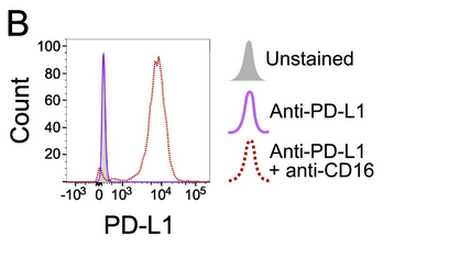
Figure 2: Staining showing that anti-PD-L1 (clone 29E.2A3) binds to AlexaFluor™ 700.
So the question comes, do we need to consider additional block to address this issue, or if we notice the issue, run an isoclonal type control, followed by a panel redesign as necessary? This is certainly one way to address the issue. In fact, Andersen comes to the rescue while discussing this issue in this recently published paper in Cytometry A. Continuing down the path of improving the quality of data and reducing or eliminating the effects of cell binding fluorochromes, the authors explored a reagent,True-Stain Blocker from Biolegend. This has been shown to minimize the binding of fluorochromes to monocytes. A second reagent, which the authors termered is ‘Oligo-Block’, or phosphorothioate‐oligodeoxynucleotides described previously in the paper by Jahrsdörfer and coworkers in 2005 to block PE-Cy5 binding. The results from the paper are shown below.
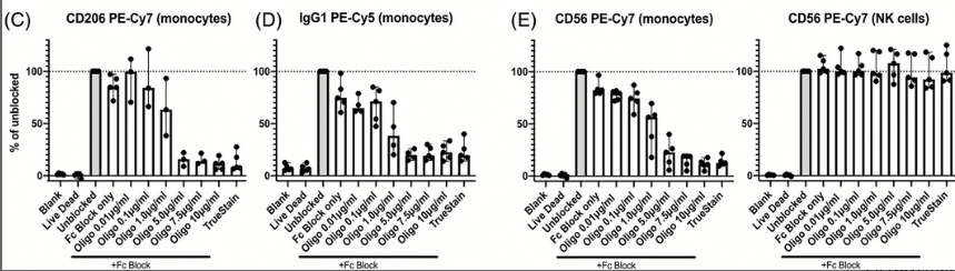
Figure 3: Effects of blocking cyanine-tandem binding on human monocytes.
Oligo-Block and True-Stain were useful in eliminating this unfortunate binding activity of monocytes, suggesting that in addition to FC block using one of these reagents is a valuable addition to your staining protocols. Getting back to the issue with the newer dyes, it turns out that Brilliant Blue 700 is a cyanine containing tandem dye.
So better safe than sorry.
Concluding Remarks
In conclusion, as you continue to optimize your flow cytometry experiments, take some time to focus on the blocking of the cells to minimize FcR mediated binding. This can be done with inexpensive human IgG serum quite easily. You can add the blocking reagent, incubate the cells for some period of time before adding the labeling reagents, and don’t have to do an extra wash, just some recalculating of the volumes.
More importantly, if you encounter some unusual staining patterns, the cause may be worth exploring. It could be the cells binding the fluorochrome, or it could be an antibody binding a fluorochrome- as shown in the PD-L1 example. Fortunately, there is a simple workaround to this, and that’s to add a second blocking reagent to minimize this effect.
It is important in the optimization phase to be extremely critical of any issues or differences you see, as you don’t want to carry something through to your production panel that could have been addressed in the optimization. Blocking is a necessary step, but often overlooked and based on lab tradition. Armed with this new knowledge, go forth and block better.
To learn more about important control measures for your flow cytometry lab, and to get access to all of our advanced materials including 20 training videos, presentations, workbooks, and private group membership, get on the Flow Cytometry Mastery Class wait list.

ABOUT TIM BUSHNELL, PHD
Tim Bushnell holds a PhD in Biology from the Rensselaer Polytechnic Institute. He is a co-founder of—and didactic mind behind—ExCyte, the world’s leading flow cytometry training company, which organization boasts a veritable library of in-the-lab resources on sequencing, microscopy, and related topics in the life sciences.
More Written by Tim Bushnell, PhD


