Understanding Reproducibility in Flow Cytometry – It’s the Antibodies!

Reproducibility is key to the scientific method. After the results of a study are published, the community validates the findings and extends them. If the findings are not reproducible, the second step is impossible. With performable experiments increasing in complexity, and the concurrent increase in the cost of equipment and reagents to perform these experiments, it is important to find the best way to maximize the money spent on advancing research.The focus of this blog entry will be on the reagents we use to identify the cells of interest: Antibodies and fluorochromes.
In flow cytometry, there are many places where improvements can be made to increase the consistency and reproducibility of an experiment. The most obvious place is in the instrument, which was the focus of a previous blog post. The focus of this blog entry will be on the reagents we use to identify the cells of interest: Antibodies and fluorochromes.
Why the focus on antibodies? The commentary on this topic (with 110 co-signers) by Bradbury and Plückthun (2015) is informative. The authors cite a review from 2008, in which the authors stated that “fewer than half of around 6,000 routinely-used commercial antibodies recognized only their specified target.” For a field that relies on these reagents on a daily basis, this is cause for some alarm.
In Begley and Ellis (2012), Begley states that in one set of experiments, only 6 of 53 “landmark preclinical studies” were able to be replicated. Bradbury and Plückthun postulate that this was “due in large part to poorly characterized antibodies.” They go on to estimate that these poorly defined reagents are costing an estimated $350 million US dollars in lost productivity in the US alone!
Baker M (2015) provided some excellent commentary on the impact it has on research when poor and uncharacterized antibodies are sold to the scientific community. The stories of David Rimm and Loannis Prassas are cautionary tales for all researchers using antibodies, as are the results published in Egelhofer et al. (2011), where 25% of 200+ tested antibodies failed specificity tests.
The most common issues to watch out for include:
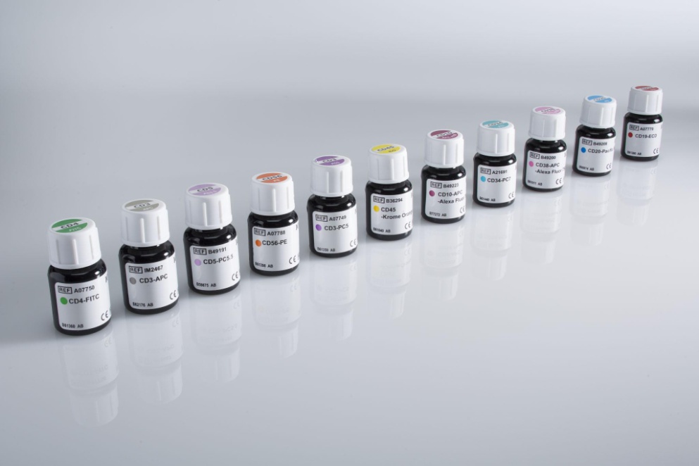
- Batch-to-batch variability of the antibodies – both the production of the antibody and its labeling with fluorochromes.
- Incorrect use of the antibody – most vendors will tell you about the applications in which their antibodies have been tested.
- Poor characterization – the antibody may have binding to secondary (off-target) proteins. This is very difficult to catch in flow cytometry, so extensive controls are necessary.
While everyone has a favorite vendor from whom they purchase common reagents, not all antibodies are created equal. It may be difficult to know whom to trust. The website Benchsci.com is a site every flow cytometrist should bookmark when searching for reagents. Benchsci’s searchable database, which contains over 3.9 million antibodies, allows researchers to narrow down species, tissue type, and even application. These can be used to identify not only antibody vendors but publications that have used the reagent in question. Recently, publishers “Springer Nature” and “Wiley” allowed Benchsci users access to figures that are normally “closed-access,” increasing the power of this tool.
Now that the problem is clear–that the reagents used in flow cytometry can negatively impact research and discovery, or worse, clinical diagnostic testing–it will be important to understand:
- How antibodies are sold and classified
- Which tests MUST be performed for each new lot of reagents
- How best to use these reagents
Before looking at the different classifications of reagents, it is important to look at a set of guidelines known as Good Manufacturing Practices, or GMP. These guidelines are overseen by governmental regulatory agencies. They are designed to ensure that products made under the GMP are consistently produced, and that the manufacturing process is rigorously controlled so that the final product meets a predefined set of quality standards.
Researchers rely on various manufacturers to produce reagents used in the laboratory. Antibodies can be purchased with several different designations, and these designations have more to do with the reagent than its cost alone. The ones most commonly used in the research lab are labeled RUO, which stands for “Research Use Only.” The RUO designation doesn’t offer a guarantee of quality, and generally, there is little effort to validate the reagent itself. In fact, when reading the technical specifications of an RUO reagent, one often finds a statement like this:
“… this product is for research use only and not intended for human or animal diagnostic, therapeutic or commercial use.”
While the vendor may do some testing and qualify the assays it will work in, there can be dramatic lot-to-lot variation using the same reagent from different batches. It is up to the vendors to decide how much to test or validate the reagent, and what they will report to the end-user; so caveat emptor applies.
The Analyte Specific Reagent designation, or ASR, is based on the US guideline 21 CFR 864.4020. These reagents must have some level of validation, and are prepared more carefully than an RUO reagent. They are intended for use in diagnostic application but must be validated in the laboratory. The FDA, the regulatory body governing these products, does not have a requirement on the performance of ASR reagents. It requires that these products carry the following on their label: “Analyte Specific Reagent. Analytical and performance characteristics are not established.”
Finally, there is IVD, or “In Vitro Diagnostic” reagents. These are usually sold as kits, and as the name implies, they are used for diagnostics. These reagents pass much stricter regulations and have been validated on actual clinical samples. Because of these regulations and validations, they are also the most expensive. Specifically, IVD reagents must meet a specific level of performance, and this must be demonstrated and documented the manufacturer.
Confused? Here is a table that summarizes the important characteristics of the differences in these three levels.
| RUO | ASR | CE-IVD | |
| Research Use | YES | YES | YES |
| Diagnostic Use | NO | YES | YES |
| FDA General Controls | YES | YES | NO |
| Manufactured under GMP | NO | YES | YES |
| Labeling requirements | YES | YES | YES |
| Performance claims | YES | N/A | YES |
| Generate data to support performance claims | YES | N/A | YES |
| Facilitated laboratory accreditation | NO | NO | YES |
| Manufacturer audit by certification body | YES | YES | YES |
Unfortunately, there are many reagents that don’t exist at the IVD level, and the ASR designation is US-centric. This means that for most work, only RUO reagents are available. In this case, it becomes important to understand the concerns over the RUO and how to use these reagents in flow cytometry experiments.
Why? Because there can be significant lot-to-lot variation between RUO reagents, and if the lab using them does not address this lot-to-lot variation, it can lead to an extensive loss of work, a lack of reproducibility, and even incorrect conclusions.
Here is the take-home message as far as the consistency and reproducibility of flow cytometry assays: Look at the manufacturing standard that the vendor is following. Vendors who follow GMP will have the best consistency across reagent lots. Clinical reagents must be manufactured under GMP; this is mandatory. While RUO reagents do not share this requirement, some vendors have made it a policy to manufacture all reagents under GMP and its strict guidelines.

FIGURE 1: “Good Manufacturing Practices” is the gold standard for ensuring high-quality reagents with lot-to-lot consistency.
In developing a polychromatic flow cytometry panel for research or clinical use, a great deal of thought needs to go into the development and pairing of fluorochrome and antibody. It is recommended that researchers and clinicians pair antigens expressed at a low level with brighter fluorochromes. They should also take into account any sensitivity-reducing issues brought about by the other fluorochromes in the panel. When determining brightness, the concept of the staining index is useful. As described in Maecker et al. (2004), the same antibody, conjugated to different fluorochromes, is used to label cells. After acquisition, the Staining Index (SI) is calculated. This is shown in Figure 2.

Figure 2: Calculating the Staining Index, as described by Maecker et al. (2004). The distance is the difference between the mean of the positive minus the mean of the negative. This is then divided by two times the spread of the negative, as measured by the standard deviation.
Of course, titration is a critical parameter for validating that the reagent is working, and that the correct staining concentration is being used. Too much antibody and there is a loss of signal due to non-specific binding of the reagent. Too little antibody and the signal decreases. An excellent review of this, and other issues that reduce sensitivity and impact the correct interpretation of data, can be found in Hulspar et al. (2006).
One other critical process for using antibodies to ensure high-quality data is the concept of the “master mix,” or antibody cocktails. This process is a very powerful tool for ensuring that each tube gets the correct about of antibody, and it saves on errors caused by pipetting small volumes.

Figure 3: The impact of not using an antibody cocktail when staining samples. Sample 1 was stained with an antibody mix made on the fly, while Sample 2 and 3 were run from an antibody cocktail.
There are those who worry about antibody cocktails for staining, especially when staining using tandem dyes, which are known to have their own issues. In Johansson and Maecy (2014), the authors addressed this issue and demonstrated the stability of different tandem dyes within cocktails up through 8 weeks. The authors also evaluated the lot-to-lot variation between different tandem dyes, and recommend that each lot of tandem dyes be characterized to determine how similar or different they are from previously used reagents.
As Bernard of Chartres wrote in the 12th century, “nanos gigantum humeris insidentes,” which was later popularized by Sir Isaac Newton as, “If I have seen further, it is by standing on the shoulders of Giants.” Scientific progress is made as each new set of experiments builds on the previous one to see farther, peer deeper, and understand more fully the biology that is being studied. This can only happen when the earlier results are robust and reproducible. The paths to cures are out there, but when only 11% of the findings from 53 preclinical papers hold up to scrutiny, that road looks longer and harder to travel than ever. Reproducibility is a mindset that every scientist must apply to their research at every step of the way, and even the littlest detail needs to be explored to ensure that published results hold up to review and scrutiny.
References
To learn more about Understanding Reproducibility in Flow Cytometry – It’s the Antibodies, and to get access to all of our advanced materials including 20 training videos, presentations, workbooks, and private group membership, get on the Flow Cytometry Mastery Class wait list.

ABOUT TIM BUSHNELL, PHD
Tim Bushnell holds a PhD in Biology from the Rensselaer Polytechnic Institute. He is a co-founder of—and didactic mind behind—ExCyte, the world’s leading flow cytometry training company, which organization boasts a veritable library of in-the-lab resources on sequencing, microscopy, and related topics in the life sciences.
More Written by Tim Bushnell, PhD


