How To Profile DNA And RNA Expression Using Next Generation Sequencing (Part-2)

In the first blog of this series, we explored the power of sequencing the genome at various levels. We also dealt with how the characterization of the RNA expression levels helps us to understand the changes at the genome level. These changes impact the downstream expression of the target genes. In this blog, we will explore how NGS sequencing can help us comprehend DNA modification that affect the expression pattern of the given genes (epigenetic profiling) as well as characterizing the DNA-protein interactions that allow for the identification of genes that may be regulated by a given protein. 

DNA Methylation Profiling or Epigenetic Profiling

NGS can be adapted to profile DNA methylation either through an enrichment (using methyl CpG antibody or methyl-CpG-binding protein) or by bisulfite sequencing.

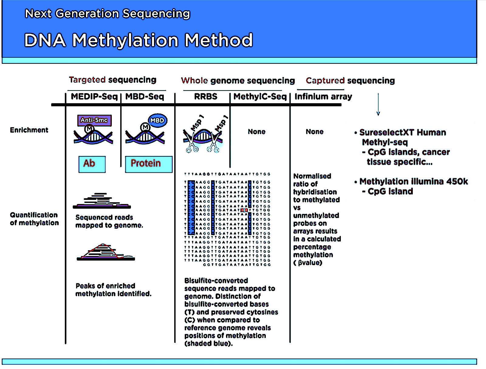
Figure 1: Different methods of NGS DNA Methylation profiling.

1. Bisulfite Sequencing

Bisulfite treatment of DNA converts unmethylated cytosines to uracil, while methylated cytosines remain the same. Uracil bases are then identified as thymine in the sequencing data, which could be used to identify the location and percentage of methylated cytosines. NGS-based bisulfite sequencing — whether whole-genome or targeted — makes it possible to profile genome-wide cytosine methylation at single-base resolution.

Types of Bisulfites sequencing:

a. Whole-Genome Bisulfite Sequencing (WGBS)

Currently, WGBS is the most comprehensive way to profile DNA methylation at base-pair resolution. However, the required depth (minimum 30x) makes it cost-prohibitive. Thus, other enrichment methods have been devised to reduce the cost of methylation profiling, especially when 100% coverage or base-pair resolution is not necessary.

b. Reduced Representation Bisulfite Sequencing (RRBS)

RRBS relies on restriction enzymes such as MspI (CCGG) or BglII (AGATCT), which tend to cut inside or near CpG islands and promoter regions regardless of methylation status. Subsequently, fragments between 40 – 220 bp are isolated and end-repaired, then treated with bisulfite and amplified with PCR. RRBS using MspI captures approximately 80% of CpG islands and 60% of promoter regions in human genomes.

2. Methylated DNA-enriched Sequencing

a. MethyCap-Seq

This sequencing uses the Methyl-CpG-binding (MBD) domain of MeCP2 to capture methylated DNA on magnetic beads. After the captured DNA is enriched with magnetic capture, the bound DNA is eluted with a high-salt solution and then used for NGS. While this is a cost-effective method, the current resolution is ~150 bp, so it is suitable for fast, large-scale, and low-resolution studies.

b. Methylated DNA Immunoprecipitation-Seq (MeDIP-Seq)

It uses an anti-methylcytosine antibody to immunoprecipitate DNA with methyl CpG. While MeDIP-Seq can be relatively inexpensive, it can yield resolutions of between 100 – 300 bp.

DNA-protein Interaction Profiling

Due to the quantitative nature of NGS, chromatin immunoprecipitation-enriched DNA can be sequenced with NGS to profile any genomic regions bound by the proteins of interest that can either be recognized with an antibody or tagged with an epitope. These include DNA-binding proteins, transcription factors, histones, histone variants, specific histone modifications, and nucleosomes.

1. ChIP-Seq (Chromatin Immunoprecipitation Sequencing)

To create a ChIP enriched library, DNA-bound proteins are cross-linked to DNA using formaldehyde, before the chromatin is cleaved. The sample is then enriched using immunoprecipitation with an antibody specific to the protein or protein modification of interest. Subsequently, the crosslinks are reversed, and then the ChIP enriched library can be assayed using quantitative PCR, microarray, or NGS. 

Difference between ChIP-chip Vs. ChIP-Seq

ChIP-chip resolution is limited by the probes’ fragment sizes on the arrays, whereas ChIP-Seq can provide single-nucleotide resolution. ChIP-Seq requires much less input DNA and provides signals with an unlimited dynamic range, depending on the sequencing depth. Additionally, ChIP-Seq makes it possible to profile repetitive regions – these are often omitted from the microarrays. Repetitive regions that are often important for epigenetic control, such as heterochromatin or microsatellites, may only be mapped with NGS.

In addition to identifying genomic regions bound by the proteins, ChIP-Seq can provide insights into the functions of the DNA-bound proteins themselves. For example, ChIP-Seq data can be used to identify the cognate binding motifs of the DNA-binding proteins. This sequence data can also be used to globally infer distances between the binding sites and genomic features, such as transcription start sites, exon-intron boundaries, 3’end of genes, and from other known binding sites.

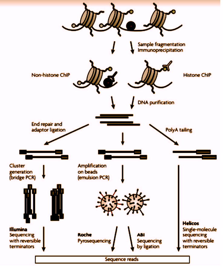
Figure 2:  A representation of Chip sequencing

  1. Micrococcal Nuclease-Seq (MNase-Seq)

Nucleosome occupancy can tell us about regions of active genes and chromatin structure in eukaryotes. NGS allows us to profile the nucleosome occupancy by sequencing the micrococcal nuclease (MNase)-digested genomic DNA. MNase prefers to digest linker DNA between histone octamers unoccupied by other proteins.

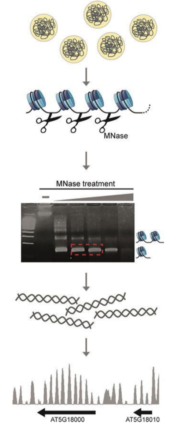
Figure 3: The workflow of an MNase protection assay

DNA is crosslinked to the protein using formaldehyde before MNase digestion. Once the digestion step is complete, the crosslinks are reversed. Then, the digested DNA is run on a gel to select the desired digested products, which are then purified and subsequently used for NGS. To control for MNase sequence bias, GC/AT preference, and other technical biases, it is necessary to concurrently sequence the genomic DNA from the same sample without crosslinking – and compare them during the analysis process.

Concluding Remarks

Over the course of these two blog posts, we have explored the power of NGS sequencing at several levels, from whole-genome sequencing, down to characterizing epigenetic differences that impact gene expression. NGS sequencing allows scientists to get a deeper holistic understanding of the genome, and variations that may be markers for the disease.  No other technique can provide such a complete picture in a relatively short time frame. As costs continue to decrease, these techniques will continue to have a greater role in areas such as drug discovery, clinical diagnostics, and ultimately personalized medicine.  Stay tuned to this blog for more information on these and many other techniques being developed in the world of NGS sequencing.   

To learn more about gene prediction and how NGS can assist you, and to get access to all of our advanced materials including 20 training videos, presentations, workbooks, and private group membership, get on the Expert Sequencing wait list.

Join Expert Cytometry's Mastery Class

ABOUT DEEPAK KUMAR, PHD

GENOMICS SOFTWARE APPLICATION ENGINEER

Deepak Kumar is a Genomics Software Application Engineer (Bioinformatics) at Agilent Technologies. He is the founder of the Expert Sequencing Program (ExSeq) at Cheeky Scientist. The ExSeq program provides a holistic understanding of the Next Generation Sequencing (NGS) field - its intricate concepts, and insights on sequenced data computational analyses. He holds diverse professional experience in Bioinformatics and computational biology and is always keen on formulating computational solutions to biological problems.

Deepak Kumar, PhD

Similar Articles

The Power Of Spectral Viewers And Their Use In Full Spectrum Flow Cytometry

The Power Of Spectral Viewers And Their Use In Full Spectrum Flow Cytometry

By: Tim Bushnell, PhD

What photon from yonder fluorochrome breaks?  It is … umm… hmmm. Let me see. Excitation off a 561 nm laser, with an emission maximum of 692 nm. I’m sure if Shakespeare was a flow cytometrist, he might have written that very scene. But the play is lost in time. However, since the protagonist had difficulty determining what fluorochrome was emitting photons, let’s consider how this could be figured out. In my opinion, one of the handiest flow cytometry tools is the spectral viewer. This tool helps visualize the excitation and emission profile of different fluorochromes, as well as allowing you…

Fickle Markers: Solutions For Antibody Binding Specificity Challenges

Fickle Markers: Solutions For Antibody Binding Specificity Challenges

By: Tim Bushnell, PhD

Reproducibility has been an ongoing, and important, concept in the sciences for years.  In the area of biomedical research, the alarm was sounded by several papers published in the early 2010’s.  Authors like Begley and Ellis, Prinz and coworkers, and Vasilevsky and colleagues, among others reported an alarming trend in the reproducibility of pre-clinical data.  These reports indicated between 50% to almost 90% of published pre-clinical data were not reproducible.  This was further highlighted in the article by Freedman and coworkers, who tried to identify and quantify the different sources of error that could be causing this crisis.  Figure 1,…

5 Common Flow Cytometry Questions, Answered

5 Common Flow Cytometry Questions, Answered

By: Tim Bushnell, PhD

I want to thank all of you who send us your questions about flow cytometry, so I thought I would dip into the old email bag and answer a few of the common ones here.  If your question isn’t answered this time, look for it to be answered in a future blog post.  Of course, if you want us to cover a specific topic, drop us a line.  1. How Fast Can I Go? This is  a common question. The allure of the ‘hi’ button is hard to resist.  The faster you go, the sooner you are finished with data…

Combining Flow Cytometry With Plant Science, Microorganisms, And The Environment

Combining Flow Cytometry With Plant Science, Microorganisms, And The Environment

By: Tim Bushnell, PhD

My first introduction to flow cytometry was talking to a professor who’d brought one on a research cruise to study phytoplankton. It was only later that I was introduced to the marvelous world that’s been my career for over 20 years.   In that time, I’ve had the opportunity to work with researchers in many different areas, exposing me to a wide variety of cell types and more important assays. What continues to amaze me is the number of different parameters we can measure, not just the number of fluorochromes, but the information we can extract from samples – animal, vegetable…

Common Numbers-Based Questions I Get As A Flow Cytometry Core Manager And How To Answer Them

Common Numbers-Based Questions I Get As A Flow Cytometry Core Manager And How To Answer Them

By: Tim Bushnell, PhD

Numbers are all around us.  My personal favorite is ≅1.618 aka ɸ aka ‘the golden ratio’.  It’s found throughout history, where it has influenced architects and artists. We see it in nature, in plants, and it is used in movies to frame shots. It can be approximated by the Fibonacci sequence (another math favorite of mine). However, I have not worked out how to apply this to flow cytometry.  That doesn’t mean numbers aren’t important in flow cytometry. They are central to everything we do, and in this blog, I’m going to flit around numbers-based questions that I have received…

3 Must-Have High-Dimensional Flow Cytometry Controls

3 Must-Have High-Dimensional Flow Cytometry Controls

By: Tim Bushnell, PhD

Developments such as the recent upgrade to the Cytobank analysis platform and the creation of new packages such as Immunocluster are reducing the computational expertise needed to work with high-dimensional flow cytometry datasets. Whether you are a researcher in academia, industry, or government, you may want to take advantage of the reduced barrier to entry to apply high-dimensional flow cytometry in your work. However, you’ll need the right experimental design to access the new transformative insights available through these approaches and avoid wasting the considerable time and money required for performing them. As with all experiments, a good design begins…

The Fluorochrome Less Excited: How To Build A Flow Cytometry Antibody Panel

The Fluorochrome Less Excited: How To Build A Flow Cytometry Antibody Panel

By: Tim Bushnell, PhD

Fluorochrome, antibodies and detectors are important. The journey of a thousand cells starts with a good fluorescent panel. The polychromatic panel is the combination of antibodies and fluorochromes. These will be used during the experiment to answer the biological question of interest. When you only need a few targets, the creation of the panel is relatively straightforward. It’s only when you start to get into more complex panels with multiple fluorochromes that overlap in excitation and emission gets more interesting.  FLUOROCHROMES Both full spectrum and traditional fluorescent flow cytometry rely on measuring the emission of the fluorochromes that are attached…

Flow Cytometry Year in Review: Key Changes To Know

Flow Cytometry Year in Review: Key Changes To Know

By: Meerambika Mishra

Here we are, at the end of an eventful year 2021. But with the promise of a new year 2022 to come. It has been a long year, filled with ups and downs. It is always good to reflect on the past year as we move to the future.  In Memoriam Sir Isaac Newton wrote “If I have seen further, it is by standing upon the shoulders of giants.” In the past year, we have lost some giants of our field including Zbigniew Darzynkiwicz, who contributed much in the areas of cell cycle analysis and apoptosis. Howard Shapiro, known for…

What Star Trek Taught Me About Flow Cytometry

What Star Trek Taught Me About Flow Cytometry

By: Tim Bushnell, PhD

It is no secret that I am a very big fan of the Star Trek franchise. There are many good episodes and lessons explored in the 813+ episodes, 12 movies (and counting). Don’t worry, this blog is not going to review all 813, or even 5 of them. Instead, some of the lessons I have taken away from the show that have applicability to science and flow cytometry.  “Darmok and Jalad at Tanagra.”  (ST:TNG season 5, episode 2) This is probably one of my favorite episodes, which involves Picard and an alien trying to establish a common ground and learn…

Top Industry Career eBooks

Get the Advanced Microscopy eBook

Get the Advanced Microscopy eBook

Heather Brown-Harding, PhD

Learn the best practices and advanced techniques across the diverse fields of microscopy, including instrumentation, experimental setup, image analysis, figure preparation, and more.

Get The Free Modern Flow Cytometry eBook

Get The Free Modern Flow Cytometry eBook

Tim Bushnell, PhD

Learn the best practices of flow cytometry experimentation, data analysis, figure preparation, antibody panel design, instrumentation and more.

Get The Free 4-10 Compensation eBook

Get The Free 4-10 Compensation eBook

Tim Bushnell, PhD

Advanced 4-10 Color Compensation, Learn strategies for designing advanced antibody compensation panels and how to use your compensation matrix to analyze your experimental data.