How small can you go? Flow cytometry of bacteria and viruses

Flow cytometers are traditionally designed for measuring particles, like beads and cells. These tend to fall in the small micron size range. Looking at the relative size of different targets of biological interest, it is clear the most common targets for flow cytometry (cells) are comparatively large (figure 1).

small
Figure 1:  Relative size of different biological targets of interest. Image modified from Bioninja.   

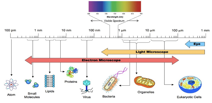
In the visible spectrum, where most of the excitation light sources reside, it is clear the cells are larger than the light. This is important as one of the characteristics that we typically measure is the amount of light that the targets scatter. This we term as either forward or side scatter, based on the position of the detector relative to the excitation light source. Forward scatter is actually the Mie scatter, first described by the German physicist Gustav Mie. Mie scatter is  the light scattered by a spherical particle that is larger than the wavelength of the light illuminating the particle. A third component impacting Mie scatter is the refractive index of the particle and media. If you are interested in exploring the impact that the wavelength and refractive index have on the Mie scatter, this calculator can be very useful.  

Even though scatter is less useful for these smaller particles, it is interesting to note that flow has been used for years to measure them. In 1979, Hercher and coworkers described the construction of a purpose built instrument capable of measuring viruses, and were able to identify different test viruses by their scatter profiles alone. A review article by McSharry titled ‘Uses of Flow Cytometry in Virology’, that was published in 1994 summarized much of the work in this field, and the possibilities have continued to expand over the years.  

Understand your instrument

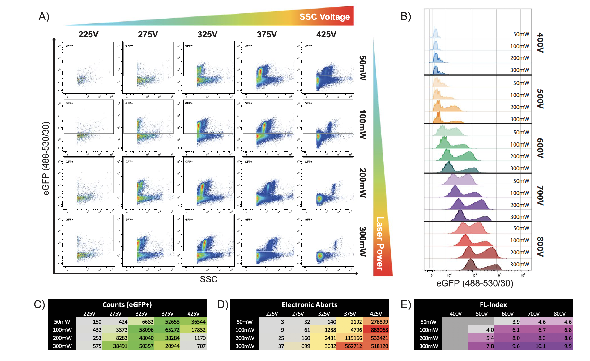
If you want  to be successful in any of these applications, the first thing that you need to understand is how sensitive your instrument is and how to get the best performance out of the system. We are fortunate that a lot of work has been done in this area because of the importance of extracellular vesicles and microparticles. In this paper by Tang et al (2017), the authors do a good job of discussing some of the work that you need to do in order to ensure that you can successfully measure smaller particles. Some of these tests include determining an appropriate threshold, how detector voltage and laser power affects the ability to resolve particles and best dilution factors to use. Take figure 2, which is taken from this paper showing one such study.  What is nice is that at the bottom there is a summary of the data looking at counts and aborts, so that it is possible to identify the best combination of voltage and laser power. 

small
Figure 2:  Effect of laser power and detector voltage on the ability to measure small particles.  

Once you have your system characterized, the fun can begin. 

Speeding up assays 

Rabies virus infected cells are detected by PCR, ELISA or electron microscopy. However, flow cytometry offers a way to potentially detect the infection of cells sooner than these techniques. Bordignon and coworkers (2002) developed an intracellular flow cytometry based assay to identify cells that were infected with the rabies virus. The literature suggested that the cells infected with rabies virus could be detected 72 hours after infection. Using the flow based assay, these researchers were able to detect virus infected cells within 12 hours post infection.  

Norden and coworkers (1995) took a similar approach to measure Mycobacterium tuberculosis by flow cytometry. In this case, they used the fact that the bacteria can hydrolyze fluorescein diacetate. The work set out to find a faster way to determine the susceptibility of M. tuberculosis to different antimicrobial agents. The standard methods took 4 to 12 days for results, and required the cells to be infected. However, the flow based assay was much faster and reduced this time to 24 hours in a cell free manner.  

Sorting bacteria and viruses

Cell sorting is an incredibly powerful tool to isolate cells. Bacteria are responsible for many processes from food production, to sewage treatment to nitrogen fixation. But many of these bacteria have never been cultured. This is where cell sorting can become highly useful, especially in marine biology. Zehr and coworkers (2008) used cell sorting to study a recently discovered single-cell nitrogen fixing cyanobacteria. This work was able to isolate small cells ( <1𝝻m in diameter) and discovered that these cells lacked the components of photosystem II, and were able to fix nitrogen during daylight hours. Since nitrogen is an element that limits plant growth, the discovery and characterization of these cells is important to better understand the overall nitrogen budget in the oceans. 

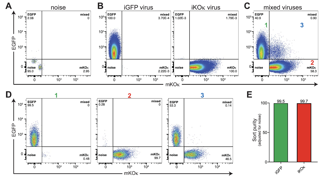
Viruses are also amenable to cell sorting, as shown in the work of Bonar and Tilton (2017). Using fluorescently labeled HIV-1 viruses, they were able to isolate  infectious HIV particles. They even performed an experiment where they mixed two different particles. One was labeled with GFP and the other with an orange emitting protein. As a proof of concept, they mixed these two viruses together and identified three populations, (top right), single positives and a double positive population. When this double positive population was sorted and reanalyzed, as expected, it was composed of the two different viral particles binding to each other as shown in the bottom panel labeled 3. 

Figure 3:  Results of viral sorting a mixture of HIV-1 labeled cells.  Taken from Bonar and Tilton (2017).

Another result from this paper demonstrated the sensitivity of flow based assays. Detection of the HIV particles was determined to be under ≤80 particles per ml. This is over 1000 times lower than the p24 ELISA and close to the sensitivity of PCR-based assays. Bonar and Tilton (2017)

Concluding Remarks

Flow cytometry is for more than just characterization and isolation of cells. Proper care and instrument characterization, enables flow cytometry to measure small particles as bacteria and viruses. Using this technique, it is possible to develop assays to speed up discovery, identify previously unknown organisms. The sensitivity of these assays as good or better than many standard assays. Perhaps it is time to consider adding flow cytometry to your experimental repertoire, if you have not already.

To learn more about important control measures for your flow cytometry lab, and to get access to all of our advanced materials including 20 training videos, presentations, workbooks, and private group membership, get on the Flow Cytometry Mastery Class wait list.

Join Expert Cytometry's Mastery Class

ABOUT TIM BUSHNELL, PHD

Tim Bushnell holds a PhD in Biology from the Rensselaer Polytechnic Institute. He is a co-founder of—and didactic mind behind—ExCyte, the world’s leading flow cytometry training company, which organization boasts a veritable library of in-the-lab resources on sequencing, microscopy, and related topics in the life sciences.

Tim Bushnell, PhD

Similar Articles

The Power Of Spectral Viewers And Their Use In Full Spectrum Flow Cytometry

The Power Of Spectral Viewers And Their Use In Full Spectrum Flow Cytometry

By: Tim Bushnell, PhD

What photon from yonder fluorochrome breaks?  It is … umm… hmmm. Let me see. Excitation off a 561 nm laser, with an emission maximum of 692 nm. I’m sure if Shakespeare was a flow cytometrist, he might have written that very scene. But the play is lost in time. However, since the protagonist had difficulty determining what fluorochrome was emitting photons, let’s consider how this could be figured out. In my opinion, one of the handiest flow cytometry tools is the spectral viewer. This tool helps visualize the excitation and emission profile of different fluorochromes, as well as allowing you…

Fickle Markers: Solutions For Antibody Binding Specificity Challenges

Fickle Markers: Solutions For Antibody Binding Specificity Challenges

By: Tim Bushnell, PhD

Reproducibility has been an ongoing, and important, concept in the sciences for years.  In the area of biomedical research, the alarm was sounded by several papers published in the early 2010’s.  Authors like Begley and Ellis, Prinz and coworkers, and Vasilevsky and colleagues, among others reported an alarming trend in the reproducibility of pre-clinical data.  These reports indicated between 50% to almost 90% of published pre-clinical data were not reproducible.  This was further highlighted in the article by Freedman and coworkers, who tried to identify and quantify the different sources of error that could be causing this crisis.  Figure 1,…

5 Common Flow Cytometry Questions, Answered

5 Common Flow Cytometry Questions, Answered

By: Tim Bushnell, PhD

I want to thank all of you who send us your questions about flow cytometry, so I thought I would dip into the old email bag and answer a few of the common ones here.  If your question isn’t answered this time, look for it to be answered in a future blog post.  Of course, if you want us to cover a specific topic, drop us a line.  1. How Fast Can I Go? This is  a common question. The allure of the ‘hi’ button is hard to resist.  The faster you go, the sooner you are finished with data…

Combining Flow Cytometry With Plant Science, Microorganisms, And The Environment

Combining Flow Cytometry With Plant Science, Microorganisms, And The Environment

By: Tim Bushnell, PhD

My first introduction to flow cytometry was talking to a professor who’d brought one on a research cruise to study phytoplankton. It was only later that I was introduced to the marvelous world that’s been my career for over 20 years.   In that time, I’ve had the opportunity to work with researchers in many different areas, exposing me to a wide variety of cell types and more important assays. What continues to amaze me is the number of different parameters we can measure, not just the number of fluorochromes, but the information we can extract from samples – animal, vegetable…

Common Numbers-Based Questions I Get As A Flow Cytometry Core Manager And How To Answer Them

Common Numbers-Based Questions I Get As A Flow Cytometry Core Manager And How To Answer Them

By: Tim Bushnell, PhD

Numbers are all around us.  My personal favorite is ≅1.618 aka ɸ aka ‘the golden ratio’.  It’s found throughout history, where it has influenced architects and artists. We see it in nature, in plants, and it is used in movies to frame shots. It can be approximated by the Fibonacci sequence (another math favorite of mine). However, I have not worked out how to apply this to flow cytometry.  That doesn’t mean numbers aren’t important in flow cytometry. They are central to everything we do, and in this blog, I’m going to flit around numbers-based questions that I have received…

3 Must-Have High-Dimensional Flow Cytometry Controls

3 Must-Have High-Dimensional Flow Cytometry Controls

By: Tim Bushnell, PhD

Developments such as the recent upgrade to the Cytobank analysis platform and the creation of new packages such as Immunocluster are reducing the computational expertise needed to work with high-dimensional flow cytometry datasets. Whether you are a researcher in academia, industry, or government, you may want to take advantage of the reduced barrier to entry to apply high-dimensional flow cytometry in your work. However, you’ll need the right experimental design to access the new transformative insights available through these approaches and avoid wasting the considerable time and money required for performing them. As with all experiments, a good design begins…

The Fluorochrome Less Excited: How To Build A Flow Cytometry Antibody Panel

The Fluorochrome Less Excited: How To Build A Flow Cytometry Antibody Panel

By: Tim Bushnell, PhD

Fluorochrome, antibodies and detectors are important. The journey of a thousand cells starts with a good fluorescent panel. The polychromatic panel is the combination of antibodies and fluorochromes. These will be used during the experiment to answer the biological question of interest. When you only need a few targets, the creation of the panel is relatively straightforward. It’s only when you start to get into more complex panels with multiple fluorochromes that overlap in excitation and emission gets more interesting.  FLUOROCHROMES Both full spectrum and traditional fluorescent flow cytometry rely on measuring the emission of the fluorochromes that are attached…

Flow Cytometry Year in Review: Key Changes To Know

Flow Cytometry Year in Review: Key Changes To Know

By: Meerambika Mishra

Here we are, at the end of an eventful year 2021. But with the promise of a new year 2022 to come. It has been a long year, filled with ups and downs. It is always good to reflect on the past year as we move to the future.  In Memoriam Sir Isaac Newton wrote “If I have seen further, it is by standing upon the shoulders of giants.” In the past year, we have lost some giants of our field including Zbigniew Darzynkiwicz, who contributed much in the areas of cell cycle analysis and apoptosis. Howard Shapiro, known for…

What Star Trek Taught Me About Flow Cytometry

What Star Trek Taught Me About Flow Cytometry

By: Tim Bushnell, PhD

It is no secret that I am a very big fan of the Star Trek franchise. There are many good episodes and lessons explored in the 813+ episodes, 12 movies (and counting). Don’t worry, this blog is not going to review all 813, or even 5 of them. Instead, some of the lessons I have taken away from the show that have applicability to science and flow cytometry.  “Darmok and Jalad at Tanagra.”  (ST:TNG season 5, episode 2) This is probably one of my favorite episodes, which involves Picard and an alien trying to establish a common ground and learn…

Top Industry Career eBooks

Get the Advanced Microscopy eBook

Get the Advanced Microscopy eBook

Heather Brown-Harding, PhD

Learn the best practices and advanced techniques across the diverse fields of microscopy, including instrumentation, experimental setup, image analysis, figure preparation, and more.

Get The Free Modern Flow Cytometry eBook

Get The Free Modern Flow Cytometry eBook

Tim Bushnell, PhD

Learn the best practices of flow cytometry experimentation, data analysis, figure preparation, antibody panel design, instrumentation and more.

Get The Free 4-10 Compensation eBook

Get The Free 4-10 Compensation eBook

Tim Bushnell, PhD

Advanced 4-10 Color Compensation, Learn strategies for designing advanced antibody compensation panels and how to use your compensation matrix to analyze your experimental data.